Muscles Of The Torso - How Many Muscles Are In The Human Body Plus A Diagram - The muscles of the human body can be categorized into a number of groups which include muscles relating to the head and neck, muscles of the torso or trunk, muscles of the upper limbs, and muscles of the lower limbs.
byAdmin-
0
Muscles Of The Torso - How Many Muscles Are In The Human Body Plus A Diagram - The muscles of the human body can be categorized into a number of groups which include muscles relating to the head and neck, muscles of the torso or trunk, muscles of the upper limbs, and muscles of the lower limbs.. Knowing which muscles do each motion will take you a long way towards proper evaluation and treatment. Media in category muscles of the human torso the following 95 files are in this category, out of 95 total. Wikimedia commons has media related to muscles of the human torso the main articles for this category are human back , human abdomen , thorax and pelvis. Mentone educational has several models that display the muscles of the torso, but one of our more extensive ones is the anatomical male muscle model. Torso spasms can sometimes be caused by simple issues like overextension of the muscles during a workout, injury to the chest wall as a result of exercise, or dehydration.
Pectoralis major (action) pectoralis minor (action) rectus abdominis (action) external obliques (action) pulls arm anteriorly and across chest, rotates humerus, and ad…. It is strapped shaped and winds across the front of the thigh, from the hip to the inner side of the tibia. When it contracts it bends and rotates the thigh. The motion of the neck can be divided into rotation (looking side to side), lateral flexion (ear to shoulder), flexion (chin to sternum) and hyperextension (looking up). This image shows the muscles of our body and displays them on both male and female diagram showing:
Images Of Torso Muscle With Label Muscles Of The Upper Torso Labeled Human Anatomy Lesson Shoulder Anatomy Shoulder Muscle Anatomy Muscle Diagram from i.pinimg.com These muscles attach the upper limb to the axial skeleton of the trunk and support the thoracic cage. The erector spinae group of muscles on each side of the vertebral column is a large muscle mass that extends from the sacrum to the skull.these muscles are primarily responsible for extending the vertebral. All of these muscles connect to the mandible and they are some of the strongest muscles in the body. Muscles of the torso diagram. Part 8 in an 8 part lecture on skeletal muscle in a flipped human anatomy course taught by wendy riggs. The muscles of the torso the trapezius is located on the back. The muscles of the human body can be categorized into a number of groups which include muscles relating to the head and neck, muscles of the torso or trunk, muscles of the upper limbs, and muscles of the lower limbs. It is strapped shaped and winds across the front of the thigh, from the hip to the inner side of the tibia.
Pages in category muscles of the torso
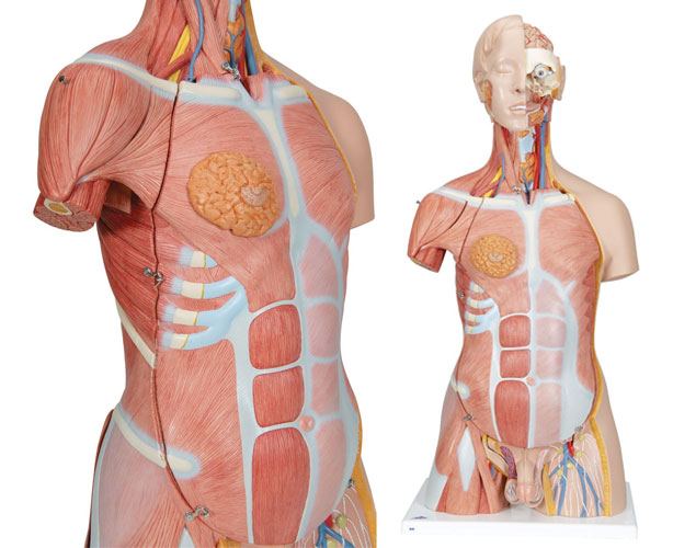
Our anatomical male muscle model displays all the deep and superficial muscles, as well as the internal organs of the human body. Media in category muscles of the human torso the following 95 files are in this category, out of 95 total. Pages in category muscles of the torso This image shows the muscles of our body and displays them on both male and female diagram showing: Injuries to this muscle are rare, but symptoms include pain in the chest, bruising, and decreased strength of the muscle. The muscles of the thoracic cage are the pectoralis major, pectoralis minor, serratus anterior, subclavius, intercostal (external, internal and innermost), subcostal and transversus thoracis muscles, including the diaphragm. On the anterior side of the thoracic region, the pectoralis minor and serratus anterior muscles originate on the anterior ribs and insert on the scapula. Mississippi gulf coast community college. Knowing which muscles do each motion will take you a long way towards proper evaluation and treatment. Mentone educational has several models that display the muscles of the torso, but one of our more extensive ones is the anatomical male muscle model. These muscles work together to move the scapula anteriorly and laterally during pushing, throwing, or punching motions. This muscle, the longest in the body, enables the crossing of the legs in the tailors's position, the function for which it is named. The muscles of the human body can be categorized into a number of groups which include muscles relating to the head and neck, muscles of the torso or trunk, muscles of the upper limbs, and muscles of the lower limbs.
The muscles of the vertebral column, thorax, and abdominal wall extend, flex, and stabilize different parts of the body's trunk. Watch the whole lecture (all 8 videos) by goin. The latissimus dorsi (dorsal) muscle is located in the lower part of the trunk. It is a complex job to balance the body on two feet and walk upright. The obliques are abdominal muscles that assist during bending and twisting of the torso.
Muscle Torso Model Life Size from www.human-anatomy.com The pectoralis minor resides under the pectoralis major. The muscles of the human body can be categorized into a number of groups which include muscles relating to the head and neck, muscles of the torso or trunk, muscles of the upper limbs, and muscles of the lower limbs. Extensor muscles of the hand 6. 'back of male torso' by thomas eakins.jpg 'front of male torso' by thomas eakins.jpg. Mississippi gulf coast community college. Knowing which muscles do each motion will take you a long way towards proper evaluation and treatment. These muscles attach the upper limb to the axial skeleton of the trunk and support the thoracic cage. Two of the muscles, temporalis and masseter, are identified in the illustration above.
The muscles of the torso the trapezius is located on the back.
These muscles attach the upper limb to the axial skeleton of the trunk and support the thoracic cage. The action refers to the action of each muscle from the standard anatomical position. The pectoralis minor is located underneath the pectoralis major. The muscles of the thoracic cage are the pectoralis major, pectoralis minor, serratus anterior, subclavius, intercostal (external, internal and innermost), subcostal and transversus thoracis muscles, including the diaphragm. Watch the whole lecture (all 8 videos) by goin. Only two of the more obvious and superficial neck muscles. Identify the movement and function of the intrinsic skeletal muscles of the back and neck, and the skeletal muscles of the abdominal wall and thorax. In this educational video children can learn about the muscles of the body and how those muscles helps us move. But muscle spasms in the torso that are felt frequently and with increased intensity can also be a sign that you're suffering from a more serious condition. Human muscles · august 21, 2016. Our anatomical male muscle model displays all the deep and superficial muscles, as well as the internal organs of the human body. Torso spasms can sometimes be caused by simple issues like overextension of the muscles during a workout, injury to the chest wall as a result of exercise, or dehydration. ( continue) describe the three types.
The quadratus lumborum muscle in the lower back side bends the lumbar spine and aids in the inspiration of air through its stabilizing affects at its insertion at the 12th rib (the last of the floating ribs). When it contracts it bends and rotates the thigh. The action refers to the action of each muscle from the standard anatomical position. Wikimedia commons has media related to muscles of the human torso the main articles for this category are human back , human abdomen , thorax and pelvis. These muscles work together to move the scapula anteriorly and laterally during pushing, throwing, or punching motions.
Dual Sex Muscle Torso Anatomy Model Deluxe 31 Parts Anatomical World Wide from d47b8c342f195720a9bf-abcdaee9f8d8752825c66eff59bb2838.ssl.cf1.rackcdn.com The erector spinae group of muscles on each side of the vertebral column is a large muscle mass that extends from the sacrum to the skull.these muscles are primarily responsible for extending the vertebral. On the anterior side of the thoracic region, the pectoralis minor and serratus anterior muscles originate on the anterior ribs and insert on the scapula. Watch the whole lecture (all 8 videos) by goin. This video covers many major muscles such as. These muscles attach the upper limb to the axial skeleton of the trunk and support the thoracic cage. Superficial and deep anterior muscles of upper body These muscles work together to move the scapula anteriorly and laterally during pushing, throwing, or punching motions. The obliques are abdominal muscles that assist during bending and twisting of the torso.
Injuries to this muscle are rare, but symptoms include pain in the chest, bruising, and decreased strength of the muscle.
The human body has three different types of muscles. Identify the movement and function of the intrinsic skeletal muscles of the back and neck, and the skeletal muscles of the abdominal wall and thorax. Human muscles · august 21, 2016. Torso spasms can sometimes be caused by simple issues like overextension of the muscles during a workout, injury to the chest wall as a result of exercise, or dehydration. This video covers many major muscles such as. The quadratus lumborum muscle in the lower back side bends the lumbar spine and aids in the inspiration of air through its stabilizing affects at its insertion at the 12th rib (the last of the floating ribs). Related posts of muscles on the side of your torso muscle anatomy lower extremity. Extensor muscles of the hand 6. The action refers to the action of each muscle from the standard anatomical position. In this educational video children can learn about the muscles of the body and how those muscles helps us move. Only two of the more obvious and superficial neck muscles. The latissimus dorsi (dorsal) muscle is located in the lower part of the trunk. The motion of the neck can be divided into rotation (looking side to side), lateral flexion (ear to shoulder), flexion (chin to sternum) and hyperextension (looking up).